Placenta Clinic Research Achievements
The research done by Dr. Kingdom and his team focuses on common pregnancy complications, including pre-eclampsia and IUGR. Below is an outline of research done at the Placenta Clinic and in Dr. Kingdom’s basic science/translational research laboratory at the Lunenfeld-Tanenbaum Research Institute.
Current understanding of antepartum haemorrhage
Dr. Kingdom is committed to advancing our understanding of pregnancy complications in the field of perinatal medicine. His recent publication is a book chapter focusing on up-to-date knowledge of antepartum haemorrhage which outlines the tools health care providers can use to ensure its proper management. To read the book chapter, click here (Baskett, Cadler, & Arulkumaran, Munro Kerr’s Operative Obstetrics, 12th Edition, Chapter 19: Antepartum Haemorrhage, 2013).
Master metabolic regulator PPAR-γ controls trophoblast differentiation
Recent work done by Khrystyna Levytska (MSc) in Dr. Kingdom’s laboratory has shown that a master metabolic regulator peroxisome proliferator-activated receptor gamma (PPAR-γ) controls the balance between cell differentiation and proliferation by regulating the expression of a downstream target glial cell missing-1 (GCM-1). Ms. Levytska’s work, based on a cellular model of trophoblast development (see Placenta 101), showed that PPAR-γ induction tips the balance toward differentiation, while blocking PPAR-γ activity has the opposite effects (click here to see the abstract). Khrystyna has presented her work at two international meetings, the International Congress on Heme Oxygenases and Related Enzymes in Edinburgh, UK (2012), and the 60th Society for Gynecologic Investigation Annual Meeting in Orlando, FL (2013).

Furthermore, Ms. Levytska’s work with Dr. Sascha Drewlo and Dora Baczyk on the role of PPAR-γ in human placentation has been presented at several meetings, notably at the Society for Gynecologic Investigation Meeting in San Diego, CA (March, 2012), where Dr. Drewlo was awarded the Pfizer President’s Achievement Award, given to best 25 abstracts out the pool of 1,200 submissions.
The emerging role of SUMOylation in placental development
Work done by Mrs. Dora Baczyk and Dr. Sascha Drewlo has resulted in findings of a novel mechanism controlling placental development. The team was first to describe the role of SUMOylation – a process involving protein modification – in healthy and pre-eclamptic placentas. As SUMOylation is also a response to cellular stress, the researchers were not surprised to find that the levels of free SUMO isoforms and SUMO-conjugated proteins are higher in diseased placentas compared to normal age-matched controls (click here to view the abstract). Dora has presented her work on the SUMO proteins at the 60th Society for Gynecologic Investigation Annual Meeting in Orlando, FL, and International Federation of Placenta Associations Meeting in Whistler, BC, in 2013.
DREAM as a novel regulator of GCM-1 in placental development
While working on her Master’s project, Mrs. Dora Baczyk studied the role of Downstream Regulatory Element Antagonist Modulator (DREAM) in placental development. Her work showed that this regulator controls the expression of GCM-1, a differentiation marker responsible for orchestrating the switch from cytotrophoblast proliferation to differentiation into the overlying syncytium (see Placenta 101 for a review of placental structure). The calcium-dependent GCM-1 repressor DREAM was found to be increased in placentas complicated by pre-eclampsia, thereby contributing to lower levels of GCM-1 and placental pathology (click here to view the abstract). Dora’s work, building on previous work done by Dr. Kingdom’s group, has shed more light on the current understanding of molecular pathways involved in placental development, and. Dora has presented her findings at the annual International Federation of Placenta Associations Meeting in Norway in 2011.
Severe placental dysfunction leading to extreme pre-term delivery is sex-specific
Male fetuses are known to suffer from more pregnancy complications, including severe pre-eclampsia , IUGR, and preterm labour, than female fetuses. Ms. Melissa Walker, a medical student at the University of Toronto (graduation year 2014), looked at the correlation between placental pathology and fetal sex (click here to view abstract). Ms. Walker found that severe placental pathology, which often results in extreme pre-term delivery (<33 weeks), is sex-specific, such that placentas with male fetuses exhibit higher rates of chronic plasma cell deciduitis and abnormal cord insertions, but a lower rate of placental infarction, compared to those with female fetuses. These observations may aid in our understanding of possible in utero mechanisms that impact the long-term development into adulthood. This work has been presented at the Society for Gynecologic Investigation Meeting in Miami, FL (March, 2011).
DUSP9 is downregulated in severe pre-eclamptic placentas
Dr. Marie Czikk (MD) undertook a study of an enzyme called dual specificity phosphatase 9 (DUSP9) during her work as a fellow in the Maternal-Fetal Medicine program at Mount Sinai Hospital. Working with Dr. Sascha Drewlo and Dora Baczyk, under the supervision of Dr. Kingdom, Marie studied the expression of this molecule in healthy pregnancy and pregnancy complications. She found that levels of DUSP9 protein are differentially expressed in placentas complicated by IUGR and pre-eclampsia, where they are elevated in IUGR placentas, while being greatly reduced in pregnancies complicated by severe pre-eclampsia (click here to view the abstract). Furthermore, Marie’s work dwelled into possible explanations for the dramatically suppressed DUSP9 expression and found that it was not due to DUSP9 promoter hypermethylation, but possibly due to prolonged hypoxia (which is a characteristic of pre-eclampsia). Surprisingly, although this gene is located on the X-chromosome and was hypothesized to be a contributing factor to the higher incidence of adverse outcomes in male pregnancies, expression levels and promoter methylation did not differ between placentas of male and female pregnancies. Dr. Czikk, who is now staff physician at Mount Sinai Hospital, presented her findings at the International Federation of Placenta Associations Meeting in Geilo, Norway, in September of 2011.
HEPRIN trial success spearheaded by two University of Toronto medical students
Leslie Proctor (Medicine Class of 2013) and Melissa Walker (Medicine Class of 2014) preceded their entries to medical school with fellowships in the Mount Sinai Hospital Placenta Clinic with Dr. Kingdom. Both with previous MSc degrees, they launched the HEPRIN trial, recruited the patients, managed the data and assisted Dr. Kingdom in preparation of the manuscript that has been published in the Journal of Thrombosis and Hemostasis (see the abstract here).

The findings of the HEPRIN trial were presented at the Society for Gynecologic Investigation Meeting in Miami, FL (March, 2011). The work was funded by a grant from the Physician’s Services Incorporated Foundation of Ontario.
Bench-to-bedside research in Dr. Kingdom’s laboratory contributes to our understanding of heparin’s actions within the placenta
Dr. Kingdom’s research team, comprising Dr. Sascha Drewlo, Ms. Dora Baczyk and Khrystyna Levytska, at the Lunenfeld-Tanenbaum Research Institute undertook the study of the role of low molecular weight heparin in the development of placental villi. To the team’s surprise, this drug increased the synthesis and release of an anti-angiogenic protein sFLT-1 from placental villi leading to the introduction of a concept of the “sFLT-1/heparin paradox:” the drug which improves pregnancy outcomes simultaneously induces the protein partially responsible for disease manifestation (click here to see the abstract of the study). This work was presented at several local, national and international meetings, most notably, at the annual Society for Gynecologic Investigation Meeting in Orlando, FL (March, 2010). Furthermore, joint efforts of Dr. Kingdom and Dr. Drewlo have resulted into a review of the role of heparin in placental function, published in 2011 in the journal Blood (click here to view the abstract).
Screening for placental insufficiency can cut the risk of stillbirth
Mount Sinai Hospital’s team led by Drs. John Kingdom and Rory Windrim have discovered that the size of a pregnant woman’s placenta can determine whether her fetus is at a high risk of stillbirth when she is found to have a low level of PAPP-A in her blood at the first trimester screening test for Down’s syndrome.
A low PAPP-A blood level typically puts the woman into the high risk group for Down’s syndrome, causing anxiety until the results of an amniocentesis test are known; however, if an alternative (and sometimes true) underlying cause of decreased PAPP-A levels (ie, placental insufficiency) is not recognized in mid-pregnancy using ultrasound, then stillbirth could occur from lack of appropriate ultrasound monitoring.
The Placenta Clinic research discovered that if women with low PAPP-A are screened during pregnancy with a placental ultrasound, those with placental abnormalities can be monitored effectively and the risk of stillbirth can be significantly reduced by planning delivery while the developing baby is still healthy. You can read the abstract of the original article published in Ultrasound in Obstetrics & Gynecology (2009) by Dr. Leslie Proctor and Dr. John Kingdom. This work was presented by Leslie Proctor at the 2009 Society for Maternal-Fetal Medicine (SMFM) meeting in San Diego, CA.
Hypospadias in males with intrauterine growth restriction due to placental insufficiency
Hypospadia is the malformation of the male genitalia which occurs in approximately 0.3-0.4% of males. The Placenta Clinic has seen a number of cases of this condition in association with intrauterine growth restriction (IUGR) with placental insufficiency. Dr. Yoav Yinon, a fellow in Maternal-Fetal Medicine, chose to study the risk of hypospadias in male IUGR pregnancies in collaboration with Leslie Proctor and Dr. John Kingdom. He identified over 40 cases of hypospadias in severe IUGR. The key points of his research findings are that:
- The degree of hypospadias is correlated to the severity of IUGR;
- IUGR is due to placental insufficiency;
- IUGR is of the early-onset type as it is commonly found with low PAPP-A;
- A high proportion of affected pregnancies were conceived using assisted reproductive technologies, like IVF.
The external male genitalia (penis, scrotum, testes) form early in the first trimester (around 7-8th week of gestation). One reason why hypospadias may be occurring in male pregnancies destined to develop severe IUGR is that an early task of the placenta is to provide the male hormone testosterone to build up the external genitalia. The implications of these findings, adopted in our Placenta Clinic, are that the sex of an IUGR fetus is medically-relevant. We therefore carefully assess the external genitalia using ultrasound in male IUGR fetuses. If hypospadias is suspected we will offer a discussion session with a pediatrician before birth. If the baby’s sex is in doubt using ultrasound, then amniocentesis may be indicated to distinguish a female developing baby from a male with severe hypospadias. Click here to see the abstract.
Male sex bias in severe placental insufficiency
Several placental complications of pregnancy (see placental insufficiency) are thought to have a male bias. In his classic textbook “Hypertensive Complications of Pregnancy,” Leon Chesley noted in the 1950’s that male offspring were more common if a woman had an eclamptic convulsion (a severe, but fortunately very rare, complication of pregnancy). Since then both stillbirth and early severe IUGR have been noted to be more common if the developing fetus is male.
We have investigated this possibility using family tree analysis of women who attended the Placenta Clinic between 2002 and 2007. Across a range of complications (stillbirth, severe IUGR, severe preeclampsia, abruption) that resulted in delivery between 20-32 weeks, more than 74% were found to be male. Family tree analysis showed that this risk is extended to male, but not female, siblings of an affected male fetus. The male bias is even stronger when a maternal risk factor like chronic hypertension (high blood pressure before becoming pregnant) is excluded.

There are two potential mechanisms. The first is genetic and may be on the X chromosome. Since a male fetus has only one X chromosome, if genes on this chromosome are defective, there is no back-up copy to work, compared to a female fetus that has an additional X chromosome (as it is XX, not XY). A number of genes and even a candidate region containing a cluster of genes on the X chromosome are needed for normal placental function. The second cause has a similar effect but is called epigenetic, meaning that the DNA (the genetic code) is intact and normal, but that the promoter region of the gene has been silenced, so gene transcription will be halted. A common mechanism to silence a gene is by methylation. We are presently studying the phenomenon of gene methylation in normal and abnormal placental development.
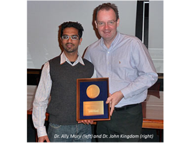
This family tree and genetic analysis work was done by Dr. Ally Murji (MD) working with Dr. Kingdom. Ally presented his work to Society for Maternal-Fetal Medicine meeting held in San Diego, CA (February, 2009). He gave a superb talk and received the prestigious Resident Research award by the Society. His findings were recently in American Journal of Medical Genetics in 2012 (click here to view the abstract).
Discovering molecular mechanisms involved in proper placental development
In 2009, molecular biology researchers Dora Baczyk (MSc) and Dr. Sascha Drewlo (PhD), working with Dr. Kingdom at the Lunenfeld-Tanenbaum Research Institute, have discovered the molecular regulator of normal placental development and trophoblast differentiation called glial cell missing-1 (GCM-1). While GCM-1 levels are deficient in placentas of severely pre-eclamptic women, they furthermore found that when GCM-1 levels are downregulated in cultured placental tissues, the placental development and differentiation is induced to resemble features found in the severely pre-eclamptic placenta (click here to see the abstract). Dora was awarded one of out of only 4 plenary awards for her work on the role of GCM-1 in placental development at the annual Society for Gynecologic Investigation Meeting in Toronto, ON (2006).

Furthermore, in March 2009, Dr. Drewlo presented these and additional findings at the annual Society for Gynecologic Investigation Meeting in Glasgow, Scotland, demonstrating that when GCM-1 is reduced in developing 1st trimester placental villi, the placental villi are induced to secrete sFLT-1 in large amounts. This led to the conclusion that reducing GCM-1 produces both the microscopic features of severe pre-eclampsia and the elevation in sFLT-1. Dr. Drewlo won a prestigious Wyeth President’s Achievement Award for his work, one of just 25 awards from over 1,200 abstracts of scientific research submitted to the meeting.
Severe early-onset pre-eclampsia is a placentally-mediated disease with a complex pathology and that gradually resolves after delivery. The central maternal feature of the disease is elevated blood pressure caused mainly by “spasm” in the arteries, termed increased systemic vascular resistance (SVR). In normal pregnancy, SVR falls to allow the healthy pregnant woman to lower her blood pressure, despite an increase in her blood volume and cardiac output. Women destined to develop severe pre-eclampsia fail to lower their blood pressure in mid-pregnancy, have poor weight gain, concentrated blood (relatively high hemoglobin levels) and then develop protein in their urine. The high SVR is considered to partially be caused by an antagonist of blood vessel relaxation, called sFLT-1. sFLT-1 is a soluble circulating receptor for vascular endothelial growth factor (VEGF), an important signal that maintains a healthy lining of blood vessels (the endothelium). The placenta destined to cause pre-eclampsia begins to release high levels of sFLT-1 several weeks before the disease of pre-eclampsia is recognized by the doctor, midwife or patient. Testing for sFLT-1 in the blood, along with another protein PlGF, is one of the new emerging tools used to screen for abnormal placental function (click here to see the abstract).
Pregnancy in the News
- Life support: Article in Science News featuring Dr. Kingdom discusses the current state of research into pregnancy complications and what can be done to help women at risk of developing them. Click here to read more!
- Bacteria linked to premature birth: Studies show that bacteria are linked to premature birth. Click here to read more!
- Babies’ weak immune systems let good bacteria in: Babies’ early lack of strong immunity lets the “good” bacteria colonize the gut. Read more here!
- Why so soon?: Scientists are trying to find the causes for premature birth and understand the mechanisms of preterm labour. Click here for an updated review of progress so far.
- Youngest preemies at high risk of neurodevelopmental problems later on: Studies assess the risk of neurological damage in premature babies. Click here to find out more.
- Older moms face more pregnancy complications: This CBC News article, quoting Dr. John Kingdom, discusses the risks associated with pregnancy in older women. Read the full story here.
